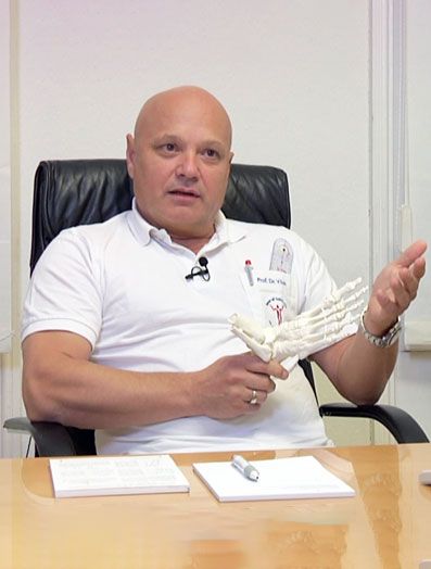
Complex deformities of the forefoot
Foot surgery without drill wires
Not infrequently, the entire foot is in a defective position.
Besides the flatfoot, other deformities occur most frequently on the forefoot.
Three examples and their correction:
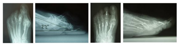
1st example:
A 72-year-old woman with massive misalignment and load-dependent ball-pain. She wants to walk and even go skiing, both not possible before the operation.
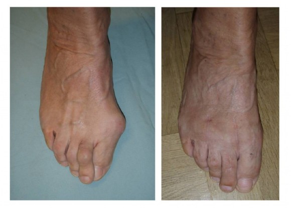
Before and 6 months after a complex forefoot correction.
The hallux was straightened and placed towards the ground, the sprained II toe repositioned and the extensor tendons lengthened, and the torn connective tissue plate at the soles was restored.
The metatarsals II, III. and IV were shortened.
The patient is able to walk and even resume sport activity.
Pain is gone.
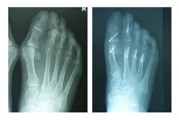
X-ray before and after the complex bone and soft tissue surgery. A metal removal is not necessary thanks to the titanium screws.
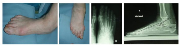
2nd example:
64-year-old woman with crossed toes and massive splayfoot problems at the ball of the foot.
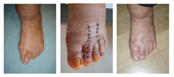
Left picture: Bad positioning of the veins and obesity aggravate the procedure.
Middle: On the third day after the operation a little swelling. The new surgical technique makes sure no wires stick out. We have applied our drill wire-free foot surgery.
Right image: routine check after 6 years. Beautiful, fully corrected foot. The patient was able to walk well a few weeks after the surgery, the swelling faded away rapidly despite the moderate initial situation.
The X-ray before the operation shows the massive abrasion of the big toe joint, the crossed II toe and the dislocation of the II and III toe joint and a subluxation of the IV toe joint.
In a side view the extent of the toe sprains can be estimated. On the right, the correction using cheilectomy at the big toe joint, which was now much more flexible, cartilage-sparing shortening of the metatarsal II, III and IV. Stiffening of the middle joint of the II toe (PIP arthrodesis) and extensive soft tissue corrections.
On the right a side view is given which shows the better situation of the toes after surgery.
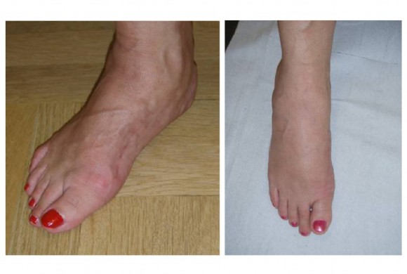
3rd example:
68-year-old woman. Acquired flatfoot due to a tendon tear at the posterior calf muscle (tibialis posterior), which is extremely essential for maintaining the arch. The patient has already undergone previous surgery elsewhere, but without success, the foot broke again and hurts with every step. In addition, the patient suffered from a hallux valgus.
The longitudinal arch is broken, the foot has no support inside. The X-ray shows impaired angles.
The surgery was performed using a 3-dimensional correction of the first metatarsal.
The longitudinal arch was restored. Between the ankle and the heel bone additionally an implant was built in to support the arch.
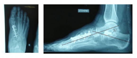
The X-ray shows a foot back in its normal position. The patient is pain-free and can walk very satisfactorily. Because of the previous surgery and the long, poor gait, walking normally again had to be practised hard, however
