Hallux valgus in adolescents
About the surgery
For the very moving feet of children and adolescents, a surgery described by McBride is known since the 1930s, which preserves the joint, and no bone cut is necessary at the first metatarsal. The tendon of the so-called adductor muscle, that pulls the big toe in the deformity, is simply transferred to the contrary now to pull the abducted first metatarsal towards the middle of the foot.
A very elegant and gentle method for the tissue. The weak point of the operation, namely the attachment of the tendon to the first metatarsal has been improved by Prof. Michael Vitek, as now a bone canal is formed in the first metatarsal. The tendon is then fed into the bone canal and firmly attached with our newly developed titanium screw.
Important: In this operation, there are no cuts made at the joints and growth areas. This does not damage the adolescent foot and growth is not affected by the operation.
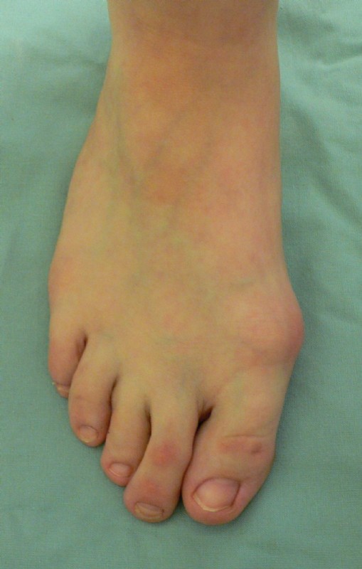
Pictures
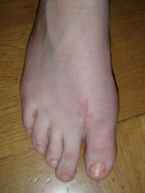
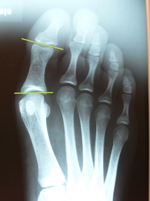
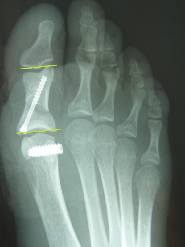
After correction the toe joints are parallel (horizontal green lines), the "ganglion" is gone, the first metatarsal is closer to the second metatarsal, and the foot ist narrower.
At the first metatarsal our new "interference screw" can be seen to have fixed the offset adductor tendon.
The toe itself is straightened by an operation according to Akin. A saw guide is designed by Prof. Dr. Michael Vitek, which enables very precise bone cuts. The fully retractable screw is also a separate new development. It allows a faster bone recovery due to a dynamic compression.
This way, only the young or the very mild Hallux deformities are treated.
For everyone else, so for about 90% of all hallux deformities, our newer, so called intramedullary locking plates are used. This is a completely new approach to hallux surgery, allowing complete correction of all deformities with immediate full activity resumable, as the plate (like a nail in trauma surgery) sits inside the bone and allows all possible correction. The forces are retained where they occur, namely in the center of the bone axis and not the outside, as has been the case with all other fixation systems so far.
